
Research regarding jaw deformities
Our classroom carries out clinical and basic research regarding jaw deformities.
CLINICAL RESEARCH
- Stability after orthognathic surgery
- Evaluation of stomatognathic functions
BASIC RESEARCH
- Elucidation of cross-bite pathogenesis using mice with spontaneous malocclusion
- Genome-wide association analysis of skeletal mandibular protrusion
CLINICAL RESEARCH
1. Stability after surgical orthodontic treatment
The Hokkaido University Hospital Dental Care Center takes a three-team approach to jaw deformity cases, where the Oral Surgery Department, the Prosthodontics Department, and the Orthodontics Department aim to carry out treatment to achieve stable desirable occlusion (D.O). Factors related to stability after jaw surgery have been investigated, and, using a variety of measures, a stable therapeutic result is achieved.
Until present, we have examined:
- Sagittal plane rotation of the lower jaw bone during orthognathic surgery
- Ensuring centric stops in posterior region after removal of intermaxillary fixation
- Harmony etc. of width and diameter of upper and lower dental arches.
2. Evaluation of stomatognathic functions
Many patients with skeletal reversed occlusion who are suitable for orthodontic surgery don’t just complain of maxillofacial morphological disharmony, but also claim abnormal oral functions such as chewing and pronunciation.
Pronunciation failure due to malocclusion is said to be particularly common with consonants such as plosives and fricatives. In our classroom, we carried out pronunciation analysis and hearing tests with /s/, the unvoiced fricative most commonly affected in the Japanese language, and discovered the acoustic features of utterances of patients with skeletal reversed occlusion, their auditory impressions, and the cause of pronunciation failure.
Currently, we are estimating the position of the articulation point (formed in the narrow portion of the oral cavity by articulation) of the 5 Japanese vowel sounds based on their acoustic characteristics. From the results, we are evaluating the difference in the position of the articulation point of each vowel between patients with skeletal reversed occlusion and healthy subjects, and the effects of orthognathic surgery.
BASIC RESEARCH
1. Elucidation of cross-bite pathogenesis using mice with spontaneous malocclusion
In orthodontics, identifying the exact pathology of malocclusion is a research topic that is related to the very foundation of orthodontic treatment. However, the difficulty in finding laboratory animals in which the state of malocclusion exists naturally has been a major constraint in research expansion until today. For these reasons, laboratory animals exhibiting a malocclusion state congenitally have been sought. It was discovered that of the BALB/c-bm/bm mice attained by transferring the bm (brachymorphism) gene from C57BL-bm/bm mice with congenital brachymelia to BALB/c mice through natural mating, about 10% developed natural anterior left and right malocclusions. With the aim of establishing the BALB/c-bm/bm mouse as a malocclusion model animal, morphological and histological examinations of the maxillofacial unit are being carried out to elucidate the malocclusion pathogenesis.
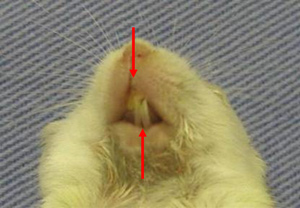
Figure 1: BALB/c-bm/bm incisor horizontal malocclusion
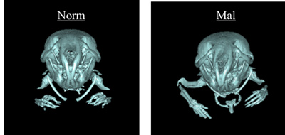
Figure 2: Craniomaxillofacial section 3DCT image of BALB/c-bm/bm normal occlusion (left) and malocclusion (right)
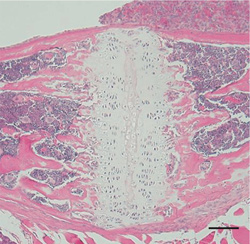
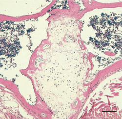
Figure 3: Image of mouse norma sagittalis, spheno-occipital synchondrosis portion
Left: BALB/c mouse, right: BALB/c-bm/bm mouse. Obvious difference in formation of cartilage.
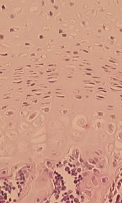
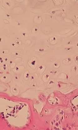
Figure 4: Image of mouse norma frontalis, septal cartilage
Left: BALB/c mouse, right: BALB/c-bm/bm
2. Genome-wide association analysis of skeletal mandibular protrusion
Skeletal mandibular protrusion is the excessive growth of the lower front jaw, and causes a feeling of significant lower jaw projection in one’s side profile and dysmasesis due to reversed occlusion. Orthognathic surgery is often a necessary treatment. It is not yet clear whether it is caused by genetic and environmental factors, but a confirmed disease-causing gene would be important in determining orthodontic treatments. There are several association analyses and relation analyses for causative gene analysis methods for skeletal mandibular protrusion as a multifactorial disease, and from several relation analyses regarding the disease, it has been suggested there is an association with the first chromosome. However, there are no reports of genome-wide association analyses regarding the disease. In this class, research regarding genome-wide association analysis (GWAS) using microsatellites is being carried out together with Tokai University School of Medicine Molecular Life Sciences Department with the aim of identifying the causative gene.
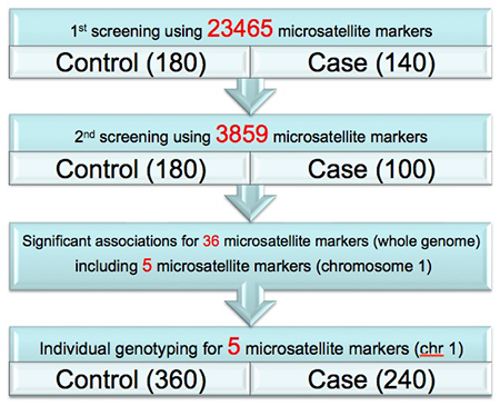
Figure 5: Narrowing down the candidate region (screening) with pooled DNA Method from markers in whole genome
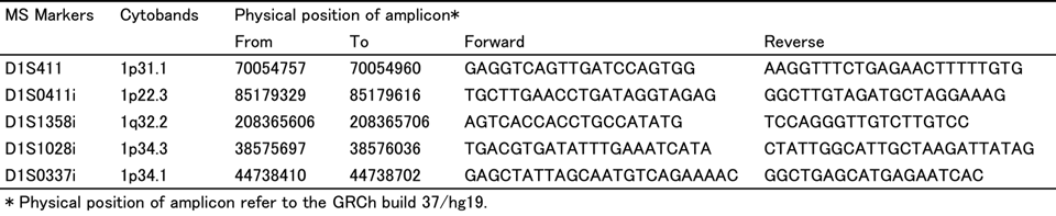
Table 1. 5 markers selected from screening prediction
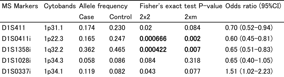
Table 2. Typing results for 5 markers
At this point, two suggested regions related to mandibular protrusion can be confirmed.
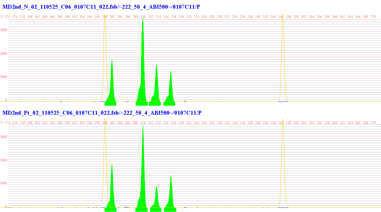
(Multi peaks, Applied Biosystems)
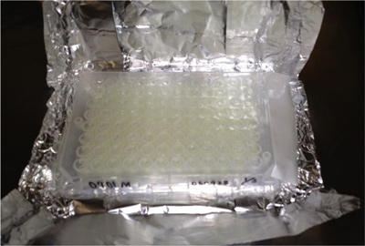
95 wells × 247 plates = 23465 markers